 Energy Dispersive X-ray Spectroscopy (EDS or EDX) is a qualitative and quantitative X-ray microanalytical technique that provides information on the chemical composition of a sample for elements with atomic number (Z) >3.
Characteristic X-ray Generation
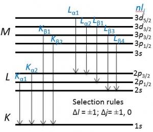
The atoms are ionized by the primary electron beam leading to holes generated on the core shells; following ionization the electrons from outer shells fill the holes and cause the emission of X-ray fluorescence lines.
The characteristic X-ray lines are named according to the shell in which the initial vacancy occurs and the shell from which an electron drops to fill that vacancy.
For instance, if the initial vacancy occurs in the K shell and the vacancy filling electron drops from the adjacent (L) shell, a Kα x-ray is emitted. If the electron drops from the M shell (two shells away), the emitted x-ray is a Kβ x-ray. Similarly, if an L-shell electron is ejected and an electron from the M-shell fills the vacancy, Lα radiation will be emitted.
EDS Detector
The detector is based on a semiconductor device, usually a crystal of silicon. The first detector developed was the lithium-drifted silicon or Si(Li) detector, which is now giving way to the 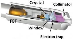 silicon-drift detector or SDD.
A typical EDS detector is composed of
- A collimator to ensure that only X-rays generated from where the primary electron beam interacts with the sample will be collected.
- An electron trap to ensure that X-rays, but no electrons, enter the detector.
-
A window to isolate the detector crystal, under high vacuum, from the chamber of the microscope. Older windows were composed of Be which did not allow low-energy X-rays (< ~0.9 keV) to pass through it, but more modern windows are composed of polymers that will allow low-energy X-rays (down to ~0.1 keV) to pass.
-
A semiconductor crystal detector.
-
Electronics to detect the charge recorded by the detector, convert it to a voltage pulse and pass it to the pulse processor.
Detector Operating Principle
- The energy of the incoming X-ray is dissipated by the creation of a series of electron-hole pairs in the semiconductor crystal.
- A high bias voltage is applied across the crystal and this causes electrons and holes to move to electrodes on opposite sides of the crystal, producing a charge signal which is passed to the pulse processor.
- The size of the signal is proportional to the energy of the incoming X-ray. For a silicon detector, ~3.8 eV is used to generate each electron-hole pair (~2.9 eV for Ge). So for an incoming Ni Kα X-ray of energy 7.477 keV, 1968 electron-hole pairs will be produced, and for an Al Kα X-ray of 1.487 keV, 391 electron-hole pairs will be generated.
- By measuring the amount of current produced by each X-ray photon, the original energy of the X-ray can be calculated. An EDS spectrum is essentially a histogram of the number of X-rays measured at each energy.